DETAIL
■この商品は「予約販売商品」です
・この商品はコンビニ払いがご利用できません。
・Erler-Zimmer GmbH & Co.KG(ドイツ)で受注生産のため、納期は4〜6カ月かかります。
・ご注文いただいた後、入荷時期が確定いたしましたら、正式にメールにてご連絡申し上げます。
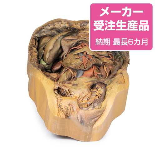
鼠径部にはヘルニアを観察できます。
Product information "Abdomen with inguinal hernia"
Diaphragm and Xyphoid Process
The diaphragm has been secured to the superior border of the dissected specimen with sutures to ensure an unobstructed view of the abdomen. The xyphoid process is in the middle of this sutured border.
Liver and Gallbladder
The liver in the right hypochondrium has been pushed laterally to reveal the kidney posterior to it.
The falciform ligament divides the right and left anatomical lobes of the liver and enveloping ligamentum teres, which is a remnant of the umbilical vein which is present during foetal development.
Below ligamentum teres at the inferior border of the liver in this model, the gall bladder is sandwiched between the anatomical lobes of the liver.
Stomach and Splenic Vasculature
The deflated stomach has been deflected superiorly to reveal the splenic artery and vein.
The tortuous course of the splenic artery and vein can be observed as it approaches the spleen, giving off numerous branches which enter the hilum of the spleen.
Spleen and Pancreas
The spleen is in the left hypochondrium of the specimen. Its gastric impression indicates where the greater curvature of the stomach would normally sit.
Toward the inferior pole of the spleen, the tail of the pancreas is fused to the hilum of the spleen. Unlike the remainder of the organ, the tail of the pancreas is intraperitoneal.
Kidneys
The kidneys are primarily retroperitoneal, however in this specimen the peritoneum that usually covers these organs has been removed.
Normally, the right kidney is displaced inferiorly by the liver relative to the than the left kidney. However, in this specimen the right kidney is both higher and smaller than the left.
The left kidney is abnormally large and is supplied by two accessory renal arteries which arise directly from the abdominal aorta. These connect just superior to the hilum and also to the inferior pole of the kidney.
Adrenal Glands
The left adrenal gland is detached from its usual position on the superior pole of the kidney. The middle adrenal artery originates directly from the aorta, left of the coeliac trunk, whilst the inferior adrenal artery is derived from the left renal artery: both supply the adrenal gland. The superior adrenal artery has been obscured by connective tissue.
Rectum and Bladder
Although the majority of the peritoneum in the abdomen has been removed below the level of the sacral prominence (S1), a layer of peritoneum remains intact, which overlays the rectum and bladder. Notably this the first part of the rectum, which is intraperitoneal.
Gastrointestinal Tract
The final part of the ascending duodenum and the descending colon at the left colic flexure has been ligated with twine, with the intestine in between being removed in order to provide a better view of the abdomen.
Pelvic Region
In this specimen the sigmoid colon has indirectly herniated through the inguinal canal.
On the right, the vas deferens emerges from the superficial inguinal ring and coursing towards the right of the scrotum to eventually attach to the right testicle. The remainder of the contents of the right spermatic cord have been removed from this specimen.
The sutures observed inferior to the vas deferens are remnants from the embalming process. These indicate that the right femoral artery was used as the point of entry.
Abdominal Vasculature
The coeliac trunk can be seen just inferior to the reflected stomach.
Typically, the coeliac trunk has three main branches; left gastric, splenic and common hepatic, to supply the foregut.
However, in this 3D model, the coeliac trunk gives off both right and left gastric branches, the splenic artery and a gastroduodenal branch that splits to become two superior pancreaticoduodenal arteries. The proper hepatic artery emerges directly from the abdominal aorta, independent of the aforementioned branches, and gives rise to the right inferior phrenic artery.
The iliolumbar artery can be seen emerging deep to the right psoas, anastomosing with branches of the right deep circumflex iliac artery which courses along the iliac crest.
●京都科学品番:EZ-160 ●メーカー品番:MP1133
Erler-Zimmer GmbH & Co.KG の模型製品は、
日本国内において株式会社京都科学の独占販売製品です。