DETAIL
■この商品は「予約販売商品」です
・この商品はコンビニ払いがご利用できません。
・Erler-Zimmer GmbH & Co.KG(ドイツ)で受注生産のため、納期は4〜6カ月かかります。
・ご注文いただいた後、入荷時期が確定いたしましたら、正式にメールにてご連絡申し上げます。
本製品の説明は英語のみです。
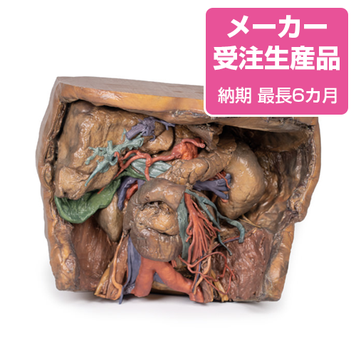
Product information "Abdomen vasculature"
Coeliac Trunk
Supplying the embryological foregut, the celiac trunk arises from T12 spinal level. Branches that can be observed in this specimen include the Left gastric artery arising from the left portion of the celiac trunk; remains of the splenic artery arising from the celiac trunk and visible passing to the left hypochondrium; the Common hepatic artery, located to the right of the celiac trunk and giving off key branches; the Gastroduodenal artery, branching inferior to into the right gastric artery, and provide an anastomosis to the superior mesentery artery via the superior pancreaticoduodenal and the Proper hepatic artery, beginning after the gastroduodenal artery, branching to form the Left hepatic artery, the first branch of the proper hepatic artery, Right hepatic artery, located inferiorly, eventually giving rise to the Cystic artery, connecting to the gallbladder.
Superior Mesenteric Artery and Inferior Mesenteric Artery
Supplying the midgut and hindgut respectively, the superior mesenteric and inferior mesenteric artery arise at the L1 and L3 vertebral levels, respectively.
While both have key branches, this specimen does not preserve them in their entirety. The Superior mesenteric artery can be seen in the model exiting below the pancreas, dividing out into many branches and the Inferior mesenteric artery can be observed descending on the left of the abdominal aorta. The left colic artery, moving laterally, can be seen leaving the IMA to give rise to the marginal arteries that supply to the colon.
Venous System of the Abdomen
The superior mesenteric vein can be seen posterior to the superior mesenteric artery, notably less tubular than its arterial counterpart.
In the specimen, the left anatomical lobe of the liver has been removed, exposing portal vein branches. These will supply nutrients from the gastrointestinal system to the hepatocytes which will then connect back to the venous system through the hepatic veins. This will then meet the Inferior Vena Cava.
Hilum of the Kidney
The right kidney shows typical anatomy, as opposed to the left kidney which shows anatomical variation. Seen at the right kidney are the Right renal vein, most superior, merging directly into the IVC, the Right renal artery, most inferior, passing deep to the IVC from its origin from the abdominal aorta and the Right ureter, coursing superficial to the right renal artery to eventually travel inferiorly.The left kidney presents unique variation at the hilum with key structures as follows. The Left renal vein, most inferior (as opposed to the usual superior) and is highly subdivided. The Left renal artery, most superior (as opposed to the usual inferior) and the Left ureter, can be seen descending from the hilum and medial to the kidney.
Muscles, Nerves and Other Vasculature
The psoas major and iliacus muscle can be seen on both sides of the specimen and surrounding them, key branches of the lumbar plexus can be seen, particularly on the left side. The Iliohypogastric nerve, continuing laterally as the most superior of the nerves present and the Ilioinguinal nerve, inferior to the iliohypogastric, directed towards the inguinal canal. The Femoral nerve, originating deep to and entering view lateral to psoas major and the Genitofemoral nerve, coursing superficial to psoas major, dividing into the genital and femoral branches of innervation.
Medial to the psoas major, the left testicular artery and left testicular vein can be seen (as this is a male specimen). While the artery will receive blood directly from the aorta, the left testicular vein will drain to the left renal vein.
The right sided testicular vasculature can also be observed however the right testicular vein drains directly into the IVC.
The branch of the iliolumbar artery that anastomoses with the iliac circumflex artery can be observed passing under the testicular artery and vein and under the ureter.
Gallbladder
Just inferior to the liver, the gallbladder can be observed with the cystic artery moving inferiorly to meet it. The cystic duct can also be seen moving from the gallbladder, meeting the common hepatic duct moving from the liver to form the common bile duct.
●京都科学品番:EZ-158 ●メーカー品番:MP1131
Erler-Zimmer GmbH & Co.KG の模型製品は、
日本国内において株式会社京都科学の独占販売製品です。